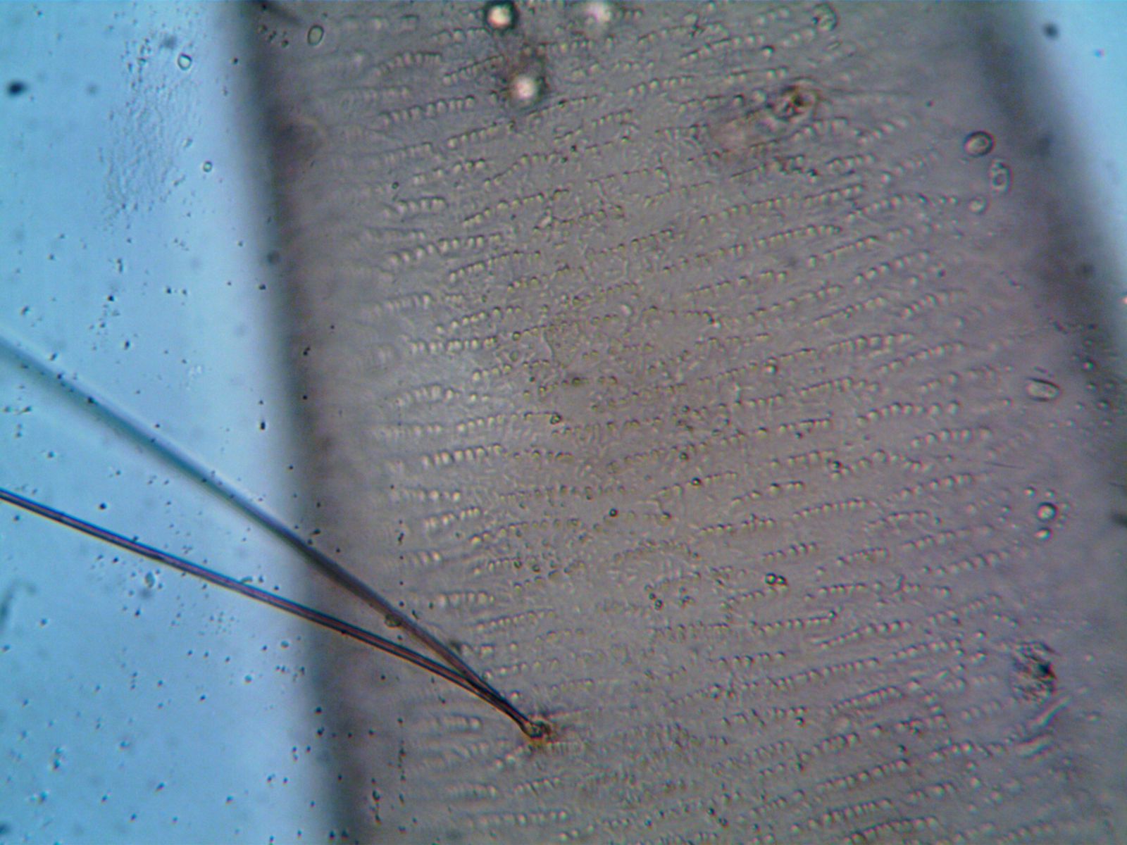
In 1665, a curly-haired natural philosopher named Robert Hooke shaved a translucent sliver from a piece of bottle cork, pressed it under the lens of a compound microscope he had built himself, and saw a honeycomb of empty rectangular chambers staring back. He wrote in his notebook that they looked like the small bare rooms monks lived in. The Latin word for those rooms was cellula. Biology got its most fundamental noun that afternoon, and it would take another 174 years before cell theory was formally established.
The observation appeared in Micrographia, published in 1665 by the Royal Society — a scientific bestseller, and a book that changed what humans thought their bodies were made of.
A slice of cork, a bad lamp, and a monastery metaphor
Cork is the outer bark of the cork oak. By the time it reaches a bottle stopper, the living tissue inside each chamber has long since died and dried away, leaving only the woody box. That is why Hooke saw compartments and not contents. He was looking at cell walls without cells.
He was working by candlelight, funnelled through a water-filled glass sphere that acted as a condenser. His microscope was a leather-and-cardboard tube about six inches long, with a small ocular lens, a field lens in the middle, and an objective at the far end. It magnified roughly 50 times. The instrument survives in reproductions — the original is lost — and a working analysis of its design reconstructs how he coaxed sharp images from ground glass and whale oil.
The word choice is worth pausing on. Hooke could have called them boxes, pores, bubbles, honeycombs. He picked the monastic image because the rows were regular, empty, and small. In doing so, he handed biology a metaphor of enclosure that survived every revision of what a cell actually is.
Who Hooke was, and why almost nobody knows his face
Robert Hooke was born on the Isle of Wight in 1635, the son of a curate. He arrived in London a sickly, spine-curved teenager with a talent for drawing and a habit of building his own instruments. By his twenties he was Robert Boyle’s paid assistant, building the air pumps that produced the first vacuum experiments. By the early 1660s he was Curator of Experiments at the Royal Society, demonstrating original experiments in front of the fellows.
Contemporaries described him as short, stooped, sharp-tongued, and preposterously productive. He surveyed London after the Great Fire of 1666. He designed buildings. He argued for an inverse-square law of gravitation years before Newton published one. He proposed that fossils were the remains of extinct animals when most scholars still called them tricks of nature.
And yet, as the plant cell biologist Larry Griffing pointed out in his investigation into the missing portrait of Hooke, there is no confirmed contemporary painting of him. Every other founding fellow of the Royal Society has a face attached to his name. Hooke has an engraving of a piece of cork.
Griffing’s forensic case is that Mary Beale’s Portrait of a Mathematician, painted in the 1680s, is Hooke — the sitter’s grey eyes, brown curls, absence of a periwig, and the elliptical-orbit diagram on the table all match Hooke’s known features and a manuscript he wrote in 1685. In a longer account of the detective work, Griffing suggests the portrait vanished from the Royal Society after Newton became its president in 1703, the year Hooke died.
Micrographia was not really a science book. It was a spectacle.
The volume is folio-sized, printed on thick paper, and contains fold-out engravings of a flea the size of a dinner plate, a louse gripping a human hair, the compound eye of a grey drone fly, the point of a needle, the edge of a razor. Samuel Pepys wrote in his diary that it was “the most ingenious book that ever I read in my life.”
The cork drawing appears as one of the volume’s observations. Hooke cut the cork two ways — across the grain and along it — and drew both, showing that the little rooms had direction. He then examined the pith of elder, carrot, fennel, and burdock, and found the same architecture. He estimated that a single cubic inch of cork contained over a billion of the little boxes.
The engravings were done by Hooke himself. He was an accomplished draughtsman, and the plates are still reproduced today. A modern gallery of cell micrographs notes that Hooke was the first to illustrate the building blocks of life as well as the first to name them, and that his drawings have never really been out of print.

Why it took so long to figure out what a living cell contained
Hooke saw empty boxes because his cork was dead. When Antoni van Leeuwenhoek, a Delft draper, built simple single-lens microscopes in the following decade, he achieved much higher magnification and began finding “animalcules” swimming in pond water, in scrapings from his own teeth, in rainwater from a barrel. Those were live cells — bacteria, protozoa, spermatozoa — but the connection to Hooke’s compartments was not made. The word cell stayed attached to the wall, not the swimmer inside it.
The reason the wait was so long is partly optical. Compound microscopes of the seventeenth and eighteenth centuries suffered from chromatic aberration: coloured fringes that fogged fine detail. The problem was not solved until the 1820s, when achromatic objectives were developed. Only then could biologists reliably see the internal contents of a living cell — the granular cytoplasm, the dense central nucleus.
The nucleus was first clearly described in 1831 by the Scottish botanist Robert Brown. In 1838 the German botanist Matthias Schleiden proposed that all plants were made of cells. In 1839 his colleague Theodor Schwann extended the claim to animals. That is the year the modern cell theory is dated to, and it is 174 years after Hooke’s cork.
The story got stranger still when the phosphorus-rich contents of the nucleus itself came under scrutiny. The Swiss physician Friedrich Miescher isolated a strange phosphorus-rich substance from pus-soaked bandages of wounded soldiers at a Tübingen clinic in 1869, called it nuclein, and was unknowingly holding purified DNA. Another 84 years would pass before Rosalind Franklin’s Photo 51 made the double helix visible.
The through-line runs from Hooke’s candlelit cork slice, to Miescher’s bandages, to Franklin’s X-ray plate. Each opened a compartment the previous one had left sealed.
The feud with Newton, and the lost face
Hooke’s public reputation took a battering after his death, and the man mostly responsible was Isaac Newton. The two clashed repeatedly — over the nature of light, over the inverse-square law of gravity, over who deserved credit for the mathematics of planetary orbits. Newton’s line about standing on the shoulders of giants, written in a letter to Hooke in 1676, is now often read as a barbed reference to Hooke’s stooped spine.
Hooke died in 1703. Newton became president of the Royal Society later that year. When the Society moved premises in 1710, Hooke’s portrait — if it existed — did not make the move. A version of the story, sifted in a profile calling Hooke the ‘English Leonardo’, holds that Newton either destroyed the painting or quietly failed to preserve it. There is no direct evidence, only absence.
The absence has become part of the story. A man who described a flea’s leg-hairs and the compartments inside a cork disc has no face of his own. He is the only major seventeenth-century natural philosopher for whom that is true. As one historian of geology has written, Hooke was the last of the virtuosi — the polymaths who felt entitled to work across every field at once — and the first casualty of the specialisation that followed.
From billions of boxes per cubic inch to 37.2 trillion cells per body
The obvious question is what Hooke would think of the current inventory. The portrait investigation piece mentions in passing that biology’s next mega-project is the Human Cell Atlas, an international effort to catalogue every one of the roughly 37.2 trillion cells in the human body. Each cell gets a molecular signature — a readout of which genes are active — and a three-dimensional address inside a tissue.
Textbooks used to list about 300 human cell types. The atlas has already found more, including two new retinal cells missed by a century of ophthalmic study, and a rare immune cell that produces a steroid capable of damping down the body’s own defences. One researcher can now sequence 10,000 individual cells in a day, at a cost of a few cents each. The consortium behind it includes the Sanger Institute, the Broad Institute of MIT and Harvard, and a Biohub in California funded by an initial $3 billion donation from Priscilla Chan and Mark Zuckerberg.
Every one of those 37.2 trillion cells is still called by the name Hooke gave a piece of dead bark in 1665. The instruments have improved by a factor of tens of thousands. The word has not moved.
What a piece of cork actually contains
Cork is made of cells whose walls are impregnated with suberin, a waxy polymer that makes the tissue waterproof and buoyant. When a cork oak in the Alentejo region of Portugal is stripped of its bark every nine years, the tree survives and regrows. A single tree can be harvested for two centuries.
The chambers Hooke drew are still there in the stopper of every wine bottle. They are the same shape, roughly rectangular, packed in offset rows. They are what makes cork springy — trapped gas inside sealed walls compresses and rebounds. Every time a cork is squeezed into a bottle neck, several billion of Hooke’s little rooms are being crushed and released.
Somewhere in a private collection, if Griffing is right, there is a canvas painted by Mary Beale showing a curly-haired man in a dark mantle, pointing at a diagram of an elliptical orbit, with a prototype orrery on the table beside him. The buildings in the background are ones he designed. He is looking directly at the viewer, and he has been waiting three hundred and twenty years for someone to look back.